Cross Section Of an Animal Cell Biography
Source:- Google.com.pkIntroduction to Animals
Sponges, Cnidarians
Characteristics of Animals
Animals are multicellular
Except for sponges, animal cells are arranged into tissues. Tissues are necessary to produce organs and organ systems.
Tissues, organs, and organ systems enabled the evolution of large, multicellular bodies.
Animal cells lack cell walls
The cells are held together by protein structures called junctions that extend from one cell to another. An abundance of extracellular proteins also support the cells.
A skeleton supports the tissues of large animals.
Animals have a period of embryonic development
During embryonic development, cells become specialized and tissues form. The growth of tissues, organs, and organ systems therefore requires a period of embryonic development.
Animals are heterotrophs
Heterotrophs consume their organic food. Except for sponges, they ingest food and digest it in a central cavity.
Recall that fungi are also heterotrophs but fungi do not ingest their food. Fungi secrete enzymes into their environment and absorb broken down organic food products.
Animals are motile
Heterotrophy often requires motility to capture prey. Animals have motility during at least some part of their life cycle.
Animals have nervous and muscle tissue
Muscle tissue allows animals to move. Nervous tissue allows rapid intercellular communication and enables coordinated movement and response to stimuli.
Animals are diploid (diplontic life cycle)
Their gametes are heterogametes (different sizes); eggs are larger than sperm.
The sperm are flagellated.
Gametes are produced by meiosis.
The development of some animals includes one or more larval stages. Larvae refers to immature individuals of species in which the body form of the immature individuals (the larvae) is very different than the body form of the adult. Because larvae and adults have different forms, they often eat different food and may live in different habitats. Larvae are transformed into adults by a developmental process called metamorphosis.
A typical animal life cycle is shown below.
Symmetry
Types of Symmetry
Radial Symmetry
The body parts of a radially symmetrical animal are arranged around a central axis so that each part extends from the center. The animal can be cut along the axis in more than one plane to produce identical halves. Animals that exhibit radial symmetry tend to be sessile (immobile). Radial symmetry allows them to reach out in all directions.
Bilateral Symmetry
Only one cut along the longitudinal axis will produce identical halves of a bilaterally symmetrical animal. Bilateral symmetry is best for motile animals.
Asymmetry
Asymmetrical animals have no pattern of symmetry. The simplest animals (sponges) are asymmetrical.
Evolution of Symmetry
Sponges lack symmetry, and Cnidarians exhibit radial symmetry. The remainder of the phyla listed below have bilateral symmetry.
Body Plans
Embryonic Development
A fertilized animal egg divides to produce a solid ball of cells. Then, cell migration results in a hollow ball called a blastula.
Some cells of the blastula migrate inward producing a gastrula. The opening is the blastopore. The tube produced by this process will become the gut (digestive tract) of the mature animal. In species that have a separate mouth and anus, the tube will eventually extend through the length of the embryo and fuse with the opposite side. One opening will become the mouth, the other will become the anus.
In the diagram below, a circle is used to represent a blastula.
Embryonic Germ Layers
The three layers of tissues that become established during early embryonic development are called germ layers. They give rise to the body tissues. These layers are ectoderm, mesoderm, and endoderm.
The diagram below shows a cross section of an animal embryo
.
The ectoderm forms from the outer layer of cells. It gives rise to the skin and nervous system.
The cells that formed the tube-like structure in the gastrula (see the diagram above) are endoderm. These cells will form the lining of the gut and the organs derived from the gut.
Mesoderm forms between the ectoderm and endoderm. It becomes the muscles, connective tissues, skeleton, kidneys, circulatory and reproductive organs
Body Cavity
The body cavity is a fluid-filled space that separates the gut and internal organs from the rest of the body.
It isolates the internal organs from body-wall movements.
It also bathes the internal organs in a liquid through which nutrients and wastes can diffuse.
Arrangement of Ectoderm, Mesoderm, and Endoderm
An acoelomate animal does not have a body cavity.
A pseudocoelomate animal has a body cavity (called a pseudocoelom) located between endoderm and mesoderm.
The body cavity of a coelomate animal (called a coelom) is located within the mesoderm.
The mesentery holds the gut in place.
The diagram below shows the body plans for nine major phyla of animals.
Gut
The gut is the digestive tract. It enables the animal to digest food outside of the cells (extracellular digestion). In animals without a digestive tract, food items are brought into the cell for digestion (intracellular digestion).
A sac-like gut has one opening. Food enters and leaves through the same opening.
A complete gut has two openings, a mouth and an anus. It is sometimes referred to as a tube-within-a-tube.
This type of gut allows for the specialization of parts along the tube. For example, part of the gut can become specialized for food storage, other parts can become specialized for secreting digestive enzymes and other parts for absorbing nutrients.
Cross Section Of an Animal Cell Animal Cell Model Diagram Project Parts Structure Labeled Coloring and Plant Cell Organelles Cake

Cross Section Of an Animal Cell Animal Cell Model Diagram Project Parts Structure Labeled Coloring and Plant Cell Organelles Cake

Cross Section Of an Animal Cell Animal Cell Model Diagram Project Parts Structure Labeled Coloring and Plant Cell Organelles Cake
Cross Section Of an Animal Cell Animal Cell Model Diagram Project Parts Structure Labeled Coloring and Plant Cell Organelles Cake

Cross Section Of an Animal Cell Animal Cell Model Diagram Project Parts Structure Labeled Coloring and Plant Cell Organelles Cake

Cross Section Of an Animal Cell Animal Cell Model Diagram Project Parts Structure Labeled Coloring and Plant Cell Organelles Cake

Cross Section Of an Animal Cell Animal Cell Model Diagram Project Parts Structure Labeled Coloring and Plant Cell Organelles Cake

Cross Section Of an Animal Cell Animal Cell Model Diagram Project Parts Structure Labeled Coloring and Plant Cell Organelles Cake
Cross Section Of an Animal Cell Animal Cell Model Diagram Project Parts Structure Labeled Coloring and Plant Cell Organelles Cake

Cross Section Of an Animal Cell Animal Cell Model Diagram Project Parts Structure Labeled Coloring and Plant Cell Organelles Cake

Cross Section Of an Animal Cell Animal Cell Model Diagram Project Parts Structure Labeled Coloring and Plant Cell Organelles Cake
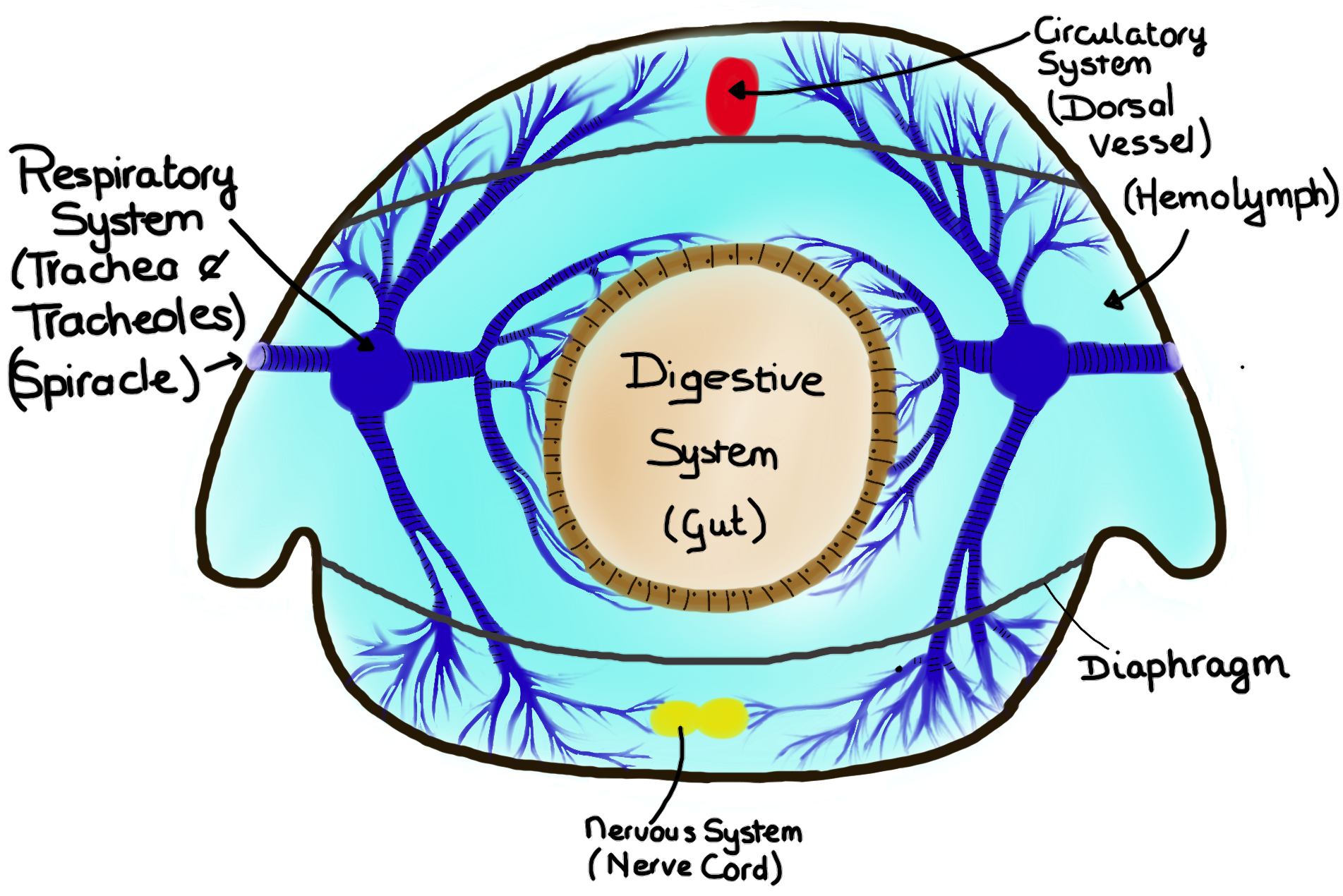
Urgent message
ReplyDeleteHi: I would like to use the image of a cross section of an insect posted in this site in a scientific paper of mine. I would use the overall aspects but not the exact colors, font and shape of the drawing (mine would be more symmetrical). Who I should ask permission to use the image in the way I have described? Thanks for your prompt reply. Sincerely, Jorge blayj@si.edu ; blayjorge@gmail.com