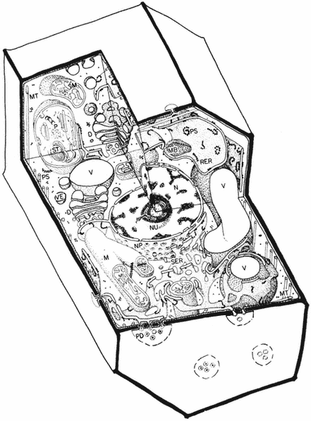
Plant And Animal Cell Diagram Biography
Source:- Google.com.pk
Today we had our second lab session. We are currently touching on the topic ‘Microscopy’ and learnt how to use the microscope today.
Oder of Events:
Newsletter activity
Strings Activity
Observing Plant and Animal Cheek Cells
Drawing Plant and Animal Cheek Cells
Newsletter Activity
In this activity, we had to choose a letter from a newspaper article and magnify it under the microscope. I chose the letter G. The result is shown below.
Bio 1Observations:
1. The letter ‘G’ is upside down
2. It is inverted
3. The paper fiber and be observed.
* This letter ‘G’ is of a magnification of 100x.
Strings
In this activity, we were supposed to magnify different coloured overlapping strings. The 3 colours I used were yellow, blue and pink.
Layout bio
The result is shown below.
Bio 2
Observations:
1. The image is inverted
2. The image is upside down.
Question: What are the thin fiber-like things?
* It is a magnification of 40x.
Observing Plant and Animal Cells
Labeling Of Animal and Plant Cells
EpitheliumEg. Cheek cell Epidermis
Squamous
1. Typical Plant Cell
Bio 4
We were given a slide of a whole mount of a plant cell. This is how the plant cell looks under the microscope.
Observations:
1. It is well structured.
2. It has an obvious nucleus
3. The cell is light blue
*It is a magnification of 100x.
The following is a picture of my friend’s (Yun Ting) observation. She managed to see the plant cell’s chloroplast under her microscope.
Bio 6
Animal Cheek Cell
Bio 5
This is a slide of a whole mount of a human cheek cell.
The human cheek cell is also know as the Human Strat Squamous Epithelium.
Observations:
1.The cell is purple in colour.
2. The cell has an irregular shape.
3. They are everywhere and are not structured.
Things to Note:
No free-hand labeling of diagrams (drawing of plant and animal cells)
No crossing lines in biology drawings
Mounted cell is called whole mount of _(eg. human cheek cell)_
Magnification must be mentioned.
We are unable to see nucleus in all plant cells because plant cells have many layers and the nucleus may be in the other layers.
Plant And Animal Cell Diagram Animal Cell Model Diagram Project Parts Structure Labeled Coloring and Plant Cell Organelles Cake


Plant And Animal Cell Diagram Animal Cell Model Diagram Project Parts Structure Labeled Coloring and Plant Cell Organelles Cake


Plant And Animal Cell Diagram Animal Cell Model Diagram Project Parts Structure Labeled Coloring and Plant Cell Organelles Cake


Plant And Animal Cell Diagram Animal Cell Model Diagram Project Parts Structure Labeled Coloring and Plant Cell Organelles Cake


Plant And Animal Cell Diagram Animal Cell Model Diagram Project Parts Structure Labeled Coloring and Plant Cell Organelles Cake


Plant And Animal Cell Diagram Animal Cell Model Diagram Project Parts Structure Labeled Coloring and Plant Cell Organelles Cake


Plant And Animal Cell Diagram Animal Cell Model Diagram Project Parts Structure Labeled Coloring and Plant Cell Organelles Cake


Plant And Animal Cell Diagram Animal Cell Model Diagram Project Parts Structure Labeled Coloring and Plant Cell Organelles Cake


Plant And Animal Cell Diagram Animal Cell Model Diagram Project Parts Structure Labeled Coloring and Plant Cell Organelles Cake


Plant And Animal Cell Diagram Animal Cell Model Diagram Project Parts Structure Labeled Coloring and Plant Cell Organelles Cake


Plant And Animal Cell Diagram Animal Cell Model Diagram Project Parts Structure Labeled Coloring and Plant Cell Organelles Cake
No comments:
Post a Comment