Diagram Of An Animal Cell Biography
Source:- Google.com.pk
2.1.1 Outline the Cell Theory
Living organisms are composed of one or more cells
Cells are the smallest unit of life
All cells come from pre-existing cells
2.1.2 Discuss the evidence for the cell theory
The cell theory has amassed tremendous credibility through the use of the microscope in the following:
Robert Hooke- studied cork and found little tiny compartments that he called cells
Antonie van Leeuwenhoek- observed the first living cells, called them 'animalcules' meaning little animals
Schleiden- stated that plants are made of 'independent, separate beings' called cells
Schwann- made a similar statement to Schleiden about animals
2.1.3 State that unicellular organisms carry out all of the functions of life
Metabolism; chemical reactions inside the cell, including cell respiration to release energy
Response; perceiving and responding to changes in the environment
Homeostasis; keeping conditions inside the organism within tolerable limits
Growth; an irreversible increase in size
Reproduction; producing offspring either sexually or asexually
Nutrition; obtaining food, to provide energy and the materials needed for growth
Defense; protection against enemies
Amoeba would be an example of an unicellular organism.
2.1.4 Compare the relative sizes of molecules, cell membrane thickness, viruses, bacteria, organelles and cells, using the appropriate SI unit
nm = nanometer µm = micrometer
Molecules - 1 nm
Thickness of membrane - 10 nm
Viruses - 100 nm
Bacteria - 1 µm
Organelles - up to 10 µm
Most cells - up to 100 µm (three dimensional nature/shape)
2.1.5 Calculate the linear magnification of drawings and the actual size of specimens in images of known magnifications
:Drawings should show cells and cell ultrastructure.
Include:
A scale bar: |------| = 1 µm
Magnification: ×250
To calculate magnification:
Magnification = Measured Size of Diagram ÷ Actual Size of Object
but before this, both magnifications must be in the same measuremente, either in mm, cm etc..
2.1.6 - Explain the importance of the surface area to volume ratio as a factor limiting cell size.
A cell needs a large surface area in order to carry out metabolic functions (as chemical reactions require a surface). As a cell grows, it needs to carry out more and more reactions. Therefore, since a cell has to maintain a certain surface area to volume ratio, its size is limited.
The rate of exchange of materials (nutrients/waste) and energy (heat) is a function of its surface area.
Thus: As a cell grows in size (volume), the distance increases between the cytoplasm at the center of the cell and the cell membrane. The rate of chemical exchange with the surrounding environment may hence become too low to maintain the cell. It is not able to excrete waste quickly enough or take in important minerals.
Volume of a cell determines requirements while surface area determines supply.
2.1.7 - State that multicelluar organisms show emergent properties
Emergent properties arise from the interaction of component parts: the whole is greater than the sum of its parts
2.1.8 - Explain how cells in multicellular organisms differentiate to carry out specialized functions by expressing some of their genes but not others.
During the early development stages of multicellular organisms, cells undergo differentiation, becoming specialized in structure and function. These cells are then organized into tissues and organs. Cells of multicellular eukaryotes express only a small fraction of their genes, allowing them to perform highly specialized functions. Cells, such as those of muscle or nervous tissue, express only a tiny fraction of their genes.
2.1.9 - State that stem cells retain the capacity to divide and have the ability to differentiate along different pathways.
Unspecialised cells that can become any type of cells.
Embryo cells are "totipotent", meaning they can become any cells; after divisions, when the zygote became a ball of cells of blastocyst, which is "pluripotent", meaning capable of being almost any type of tissue. Stem cells can also come from umbilical cord of new baby, which are "multipotent", meaning they can be limited number of tissues.
Stem cells are self-sustaining: they can divide for many times.
They differentiate into specific tissue based on a chemical signal
2.1.10 - Outline one use of therapeutic stem cells .
Bone marrow transplants. They only work because what you are actually transplanting is the hematopoetic stem cells in the marrow. And peripheral blood stem cells, as well as cord blood stem cells, can be used in lieu of bone marrow, making being a donor FAR easier today than in decades past.
http://www.marrow.org
Random Stuff that Doesn't fit or is old syllabus material
State that a virus is a non-cellular structure consisting of DNA or RNA surrounded by a protein coat.
Viruses are not cells. They are simple particles consisting of DNA and RNA wrapped in a protein coat. Viruses are not considered alive because they have no metabolism and they require a host to live. Viruses do not carry out all the functions of life, therefore they are not living.
Explain three advantages of using light microscopes.
Light microscopes
Display color instead of monochrome (black and white) images.
Provide a large field of view.
Facilitate preparation of sample material.
Allow for the examination of living material and the observation of movement.
Cheap in comparison to electron microscopes
Outline the advantages of using electron microscopes.
Electron microscopes:
Provide images of higher resolution and magnification than light microscopes.
Resolution refers to the ability to distinguish two objects as separate entities.
Magnification refers to the ability to increase the size of a viewed object.
Scanning Electron Microscopes (SEM) provide images of the specimen's surface while Transmission Electron Microscopes (TEM) provide images of a sample's interior. The resolution of an SEM is approximately half that of a TEM.
May provide a three dimensional view.
Define organelle.
An organelle is a discrete structure within a cell, and has a specific function. A mitochondrion would be an example of an organelle.
Organelle List:
mitochondrion
golgi body
endoplasmic reticulum
vacuole
lysosome
ribosome In contrast to the other organelles, they are not surrounded by a membrane.
centriole (Unique to animal cells)
chloroplast (Unique to plant cells)
Define tissue, organ and organ system.
Tissue: An integrated group of cells that share structure and are adapted to perform a similar function.
Organ: A combination of two or more tissues which function as an integrated unit, performing one or more specific functions.
Organ system: A group of organs that specialize in a certain function together.
2.2 Prokaryotic Cells[edit]
2.2.1 Draw and label a diagram of the ultrastructure of Escherichia (E. coli) as an example of a prokaryote
The Diagram Should show cell wall, plasma membrane, cytoplasm, pili, flagella, ribosomes and nuceloid (region containing naked DNA)
Two Good Pictures
http://www.the-simple-homeschool.com/image-files/prokaryote_cell_.gif
http://www.ecoliblog.com/cell-ecoli.gif
2.2.2 Annotate the diagram from 2.2.1 with the functions of each of the named structures
Cell Wall: Maintains the cell's shape and gives protection.
Plasma Membrane: Regulates the flow of materials (nutrients, waste, oxygen, etc.) into and out of the cell.
Cytoplasm: Holds and suspends the cell's specialized organelles and enzymes.
Pili: The function of the pili is attachment to solid surfaces, apparatus for use in transfer of DNA from one cell to another, twitching motility, and cell-cell adhesion.
Flagella: Flagella are whip like tails that are used to propel the organism forward.
Ribosome: Protein synthesis.
Nucleoid: Nucleoid is the area in the cytoplasm where the strands of DNA are present.
2.2.3 Identify structures from 2.2.1 in electron micrographs of E. coli
http://www.ucmp.berkeley.edu/bacteria/bacteriatem.gif http://www.exploratorium.edu/traits/images/ecoli_micro.jpg
2.2.4 State that prokaryotes divide by binary fission.
Prokaryotes divide by binary fission.
The process starts with DNA replication, then separation of the two circular strands to either side of the cell. Then cytokenesis occurs: division into two.
Each new cell has about half of the cytoplasm.
Growth restores the original size.
Old Stuff not in syllabus or misplaced
State that prokaryotes show a wide range of metabolic activity including fermentation, photosynthesis and nitrogen fixation.
Prokaryotes demonstrate a range of metabolic activity
Cyanobacteria (often referred to as blue-green algae although they are not algae) obtain their energy through photosynthesis.
Bacteria can convert organic substances into other organic substances. (i.e., glucose to lactic acid during anaerobic respiration)
Some bacteria can fix nitrogen from the air, converting it into ammonia (which is biologically available).
2.3 Eukaryotic Cells[edit]
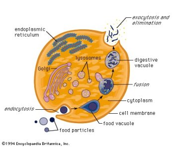
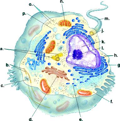
2.3.1 Draw and label a diagram of the ultrastructure of a liver cell as an example of an animal cell
Diagram of an animal cell
Should include free ribosomes, the rough endoplasmic reticulum (rER), lysosome, Golgi apparatus, mitochondrion, and nucleus.
2.3.2 Annotate the diagram from 2.3.1 with the functions of each of the named structures.
Ribosomes: Main site of protein synthesis.
Rough endoplasmic reticulum (rER): Packages the proteins synthesized in the ribosomes.
Lysosome: Digests macromolecules and contain digestive enzymes.
Golgi apparatus: Modifies, stores and routes products of the endoplasmic reticulum.
Mitochondrion: Serves as the site of cellular respiration.
Nucleus: Contains a cell's genetic material
2.3.4 Compare prokaryotic and eukaryotic cells.
Differences should include
Contain naked DNA vs. DNA associated with proteins (DNA wrapped around histones, a protein molecule, creating units called nucleosomes)
DNA in cytoplasm vs. DNA enclosed in a nuclear envelope - Prokaryotes have naked DNA in cytoplasm, Eukaryotes have a nuclear membrane surrounding it in a nucelus.
No membrane-enclosed organelles vs. membrane-enclosed organelles (e.g., mitochondria, chloroplasts) - Prokaryotes have no mitchondria, thousands of reactions occur in cytoplasm.
70S vs. 80S ribosomes - Prokaryotes = 70S, Eukaryotes = 80S.
Eukaryotic cells have internal membranes that compartmentalize their functions
prokaryotic is usually smaller in size, Eukaryotic is larger
both have cytoplasm
prokaryotic has no nucleus, Eukaryotic has a membrane-bound nucleus
prokaryotic has one chromosome / circular, Eukaryotic has two or more chromosomes
prokaryotic has DNA only, Eukaryotic has DNA with histones to bind together
prokaryotic has no membrane-bound organelles, E has some membrane-bound organelles
Eukaryotic has mitochondria, prokaryotic does not
Eukaryotic has other example of organelle, prokaryotic does not
both can have a flagellum
if flagella then E has 9+2 fibrils, prokaryotic does not
prokaryotic can have plasmids, Eukaryotic does not
both have ribosomes
prokaryotic has small ribosomes, Eukaryotic has larger ones
both have cell membrane
Eukaryotic has centriole, prokaryotic has no centriole


2.1.1 Outline the Cell Theory
Living organisms are composed of one or more cells
Cells are the smallest unit of life
All cells come from pre-existing cells
2.1.2 Discuss the evidence for the cell theory
The cell theory has amassed tremendous credibility through the use of the microscope in the following:
Robert Hooke- studied cork and found little tiny compartments that he called cells
Antonie van Leeuwenhoek- observed the first living cells, called them 'animalcules' meaning little animals
Schleiden- stated that plants are made of 'independent, separate beings' called cells
Schwann- made a similar statement to Schleiden about animals
2.1.3 State that unicellular organisms carry out all of the functions of life
Metabolism; chemical reactions inside the cell, including cell respiration to release energy
Response; perceiving and responding to changes in the environment
Homeostasis; keeping conditions inside the organism within tolerable limits
Growth; an irreversible increase in size
Reproduction; producing offspring either sexually or asexually
Nutrition; obtaining food, to provide energy and the materials needed for growth
Defense; protection against enemies
Amoeba would be an example of an unicellular organism.
2.1.4 Compare the relative sizes of molecules, cell membrane thickness, viruses, bacteria, organelles and cells, using the appropriate SI unit
nm = nanometer µm = micrometer
Molecules - 1 nm
Thickness of membrane - 10 nm
Viruses - 100 nm
Bacteria - 1 µm
Organelles - up to 10 µm
Most cells - up to 100 µm (three dimensional nature/shape)
2.1.5 Calculate the linear magnification of drawings and the actual size of specimens in images of known magnifications
:Drawings should show cells and cell ultrastructure.
Include:
A scale bar: |------| = 1 µm
Magnification: ×250
To calculate magnification:
Magnification = Measured Size of Diagram ÷ Actual Size of Object
but before this, both magnifications must be in the same measuremente, either in mm, cm etc..
2.1.6 - Explain the importance of the surface area to volume ratio as a factor limiting cell size.
A cell needs a large surface area in order to carry out metabolic functions (as chemical reactions require a surface). As a cell grows, it needs to carry out more and more reactions. Therefore, since a cell has to maintain a certain surface area to volume ratio, its size is limited.
The rate of exchange of materials (nutrients/waste) and energy (heat) is a function of its surface area.
Thus: As a cell grows in size (volume), the distance increases between the cytoplasm at the center of the cell and the cell membrane. The rate of chemical exchange with the surrounding environment may hence become too low to maintain the cell. It is not able to excrete waste quickly enough or take in important minerals.
Volume of a cell determines requirements while surface area determines supply.
2.1.7 - State that multicelluar organisms show emergent properties
Emergent properties arise from the interaction of component parts: the whole is greater than the sum of its parts
2.1.8 - Explain how cells in multicellular organisms differentiate to carry out specialized functions by expressing some of their genes but not others.
During the early development stages of multicellular organisms, cells undergo differentiation, becoming specialized in structure and function. These cells are then organized into tissues and organs. Cells of multicellular eukaryotes express only a small fraction of their genes, allowing them to perform highly specialized functions. Cells, such as those of muscle or nervous tissue, express only a tiny fraction of their genes.
2.1.9 - State that stem cells retain the capacity to divide and have the ability to differentiate along different pathways.
Unspecialised cells that can become any type of cells.
Embryo cells are "totipotent", meaning they can become any cells; after divisions, when the zygote became a ball of cells of blastocyst, which is "pluripotent", meaning capable of being almost any type of tissue. Stem cells can also come from umbilical cord of new baby, which are "multipotent", meaning they can be limited number of tissues.
Stem cells are self-sustaining: they can divide for many times.
They differentiate into specific tissue based on a chemical signal
2.1.10 - Outline one use of therapeutic stem cells .
Bone marrow transplants. They only work because what you are actually transplanting is the hematopoetic stem cells in the marrow. And peripheral blood stem cells, as well as cord blood stem cells, can be used in lieu of bone marrow, making being a donor FAR easier today than in decades past.
http://www.marrow.org
Random Stuff that Doesn't fit or is old syllabus material
State that a virus is a non-cellular structure consisting of DNA or RNA surrounded by a protein coat.
Viruses are not cells. They are simple particles consisting of DNA and RNA wrapped in a protein coat. Viruses are not considered alive because they have no metabolism and they require a host to live. Viruses do not carry out all the functions of life, therefore they are not living.
Explain three advantages of using light microscopes.
Light microscopes
Display color instead of monochrome (black and white) images.
Provide a large field of view.
Facilitate preparation of sample material.
Allow for the examination of living material and the observation of movement.
Cheap in comparison to electron microscopes
Outline the advantages of using electron microscopes.
Electron microscopes:
Provide images of higher resolution and magnification than light microscopes.
Resolution refers to the ability to distinguish two objects as separate entities.
Magnification refers to the ability to increase the size of a viewed object.
Scanning Electron Microscopes (SEM) provide images of the specimen's surface while Transmission Electron Microscopes (TEM) provide images of a sample's interior. The resolution of an SEM is approximately half that of a TEM.
May provide a three dimensional view.
Define organelle.
An organelle is a discrete structure within a cell, and has a specific function. A mitochondrion would be an example of an organelle.
Organelle List:
mitochondrion
golgi body
endoplasmic reticulum
vacuole
lysosome
ribosome In contrast to the other organelles, they are not surrounded by a membrane.
centriole (Unique to animal cells)
chloroplast (Unique to plant cells)
Define tissue, organ and organ system.
Tissue: An integrated group of cells that share structure and are adapted to perform a similar function.
Organ: A combination of two or more tissues which function as an integrated unit, performing one or more specific functions.
Organ system: A group of organs that specialize in a certain function together.
2.2 Prokaryotic Cells[edit]
2.2.1 Draw and label a diagram of the ultrastructure of Escherichia (E. coli) as an example of a prokaryote
The Diagram Should show cell wall, plasma membrane, cytoplasm, pili, flagella, ribosomes and nuceloid (region containing naked DNA)
Two Good Pictures
http://www.the-simple-homeschool.com/image-files/prokaryote_cell_.gif
http://www.ecoliblog.com/cell-ecoli.gif
2.2.2 Annotate the diagram from 2.2.1 with the functions of each of the named structures
Cell Wall: Maintains the cell's shape and gives protection.
Plasma Membrane: Regulates the flow of materials (nutrients, waste, oxygen, etc.) into and out of the cell.
Cytoplasm: Holds and suspends the cell's specialized organelles and enzymes.
Pili: The function of the pili is attachment to solid surfaces, apparatus for use in transfer of DNA from one cell to another, twitching motility, and cell-cell adhesion.
Flagella: Flagella are whip like tails that are used to propel the organism forward.
Ribosome: Protein synthesis.
Nucleoid: Nucleoid is the area in the cytoplasm where the strands of DNA are present.
2.2.3 Identify structures from 2.2.1 in electron micrographs of E. coli
http://www.ucmp.berkeley.edu/bacteria/bacteriatem.gif http://www.exploratorium.edu/traits/images/ecoli_micro.jpg
2.2.4 State that prokaryotes divide by binary fission.
Prokaryotes divide by binary fission.
The process starts with DNA replication, then separation of the two circular strands to either side of the cell. Then cytokenesis occurs: division into two.
Each new cell has about half of the cytoplasm.
Growth restores the original size.
Old Stuff not in syllabus or misplaced
State that prokaryotes show a wide range of metabolic activity including fermentation, photosynthesis and nitrogen fixation.
Prokaryotes demonstrate a range of metabolic activity
Cyanobacteria (often referred to as blue-green algae although they are not algae) obtain their energy through photosynthesis.
Bacteria can convert organic substances into other organic substances. (i.e., glucose to lactic acid during anaerobic respiration)
Some bacteria can fix nitrogen from the air, converting it into ammonia (which is biologically available).
2.3 Eukaryotic Cells[edit]
2.3.1 Draw and label a diagram of the ultrastructure of a liver cell as an example of an animal cell
Diagram of an animal cell
Should include free ribosomes, the rough endoplasmic reticulum (rER), lysosome, Golgi apparatus, mitochondrion, and nucleus.
2.3.2 Annotate the diagram from 2.3.1 with the functions of each of the named structures.
Ribosomes: Main site of protein synthesis.
Rough endoplasmic reticulum (rER): Packages the proteins synthesized in the ribosomes.
Lysosome: Digests macromolecules and contain digestive enzymes.
Golgi apparatus: Modifies, stores and routes products of the endoplasmic reticulum.
Mitochondrion: Serves as the site of cellular respiration.
Nucleus: Contains a cell's genetic material
2.3.4 Compare prokaryotic and eukaryotic cells.
Differences should include
Contain naked DNA vs. DNA associated with proteins (DNA wrapped around histones, a protein molecule, creating units called nucleosomes)
DNA in cytoplasm vs. DNA enclosed in a nuclear envelope - Prokaryotes have naked DNA in cytoplasm, Eukaryotes have a nuclear membrane surrounding it in a nucelus.
No membrane-enclosed organelles vs. membrane-enclosed organelles (e.g., mitochondria, chloroplasts) - Prokaryotes have no mitchondria, thousands of reactions occur in cytoplasm.
70S vs. 80S ribosomes - Prokaryotes = 70S, Eukaryotes = 80S.
Eukaryotic cells have internal membranes that compartmentalize their functions
prokaryotic is usually smaller in size, Eukaryotic is larger
both have cytoplasm
prokaryotic has no nucleus, Eukaryotic has a membrane-bound nucleus
prokaryotic has one chromosome / circular, Eukaryotic has two or more chromosomes
prokaryotic has DNA only, Eukaryotic has DNA with histones to bind together
prokaryotic has no membrane-bound organelles, E has some membrane-bound organelles
Eukaryotic has mitochondria, prokaryotic does not
Eukaryotic has other example of organelle, prokaryotic does not
both can have a flagellum
if flagella then E has 9+2 fibrils, prokaryotic does not
prokaryotic can have plasmids, Eukaryotic does not
both have ribosomes
prokaryotic has small ribosomes, Eukaryotic has larger ones
both have cell membrane
Eukaryotic has centriole, prokaryotic has no centriole
Diagram Of An Animal Cell Animal Cell Model Diagram Project Parts Structure Labeled Coloring and Plant Cell Organelles Cake
Diagram Of An Animal Cell Animal Cell Model Diagram Project Parts Structure Labeled Coloring and Plant Cell Organelles Cake
Diagram Of An Animal Cell Animal Cell Model Diagram Project Parts Structure Labeled Coloring and Plant Cell Organelles Cake
Diagram Of An Animal Cell Animal Cell Model Diagram Project Parts Structure Labeled Coloring and Plant Cell Organelles Cake
Diagram Of An Animal Cell Animal Cell Model Diagram Project Parts Structure Labeled Coloring and Plant Cell Organelles Cake
Diagram Of An Animal Cell Animal Cell Model Diagram Project Parts Structure Labeled Coloring and Plant Cell Organelles Cake
Diagram Of An Animal Cell Animal Cell Model Diagram Project Parts Structure Labeled Coloring and Plant Cell Organelles Cake
Diagram Of An Animal Cell Animal Cell Model Diagram Project Parts Structure Labeled Coloring and Plant Cell Organelles Cake
Diagram Of An Animal Cell Animal Cell Model Diagram Project Parts Structure Labeled Coloring and Plant Cell Organelles Cake
Diagram Of An Animal Cell Animal Cell Model Diagram Project Parts Structure Labeled Coloring and Plant Cell Organelles Cake
Diagram Of An Animal Cell Animal Cell Model Diagram Project Parts Structure Labeled Coloring and Plant Cell Organelles Cake
No comments:
Post a Comment