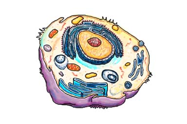
Typical Animal Cell Biography
Source:- Google.com.pk
Now, let's slog through the cytosol a bit. Notice that stack of a half dozen flattened balloons, each a few inches across and about 2 feet long? That's the Golgi complex, also called the Golgi apparatus or, simply, the Golgi. Like an upscale gift shop that monograms, wraps, and mails its merchandise, the Golgi receives newly made proteins and lipids from the ER, puts the finishing touches on them, addresses them, and sends them to their final destinations. One of the places these molecules can end up is in lysosomes.
Back to Top
Lysosomes: Recycling Centers and Garbage Trucks
Lysosomes
Lysosomes
See that bubble about 10 feet across? That's a lysosome. Let's go—I think you'll like this. Perhaps even more than other organelles, lysosomes can vary widely in size—from 5 inches to 30 feet across.
Go ahead, put your ear next to it. Hear the sizzling and gurgling? That's the sound of powerful enzymes and acids chewing to bits anything that ends up inside.
But materials aren't just melted into oblivion in the lysosome. Instead, they are precisely chipped into their component parts, almost all of which the cell recycles as nutrients or building blocks. Lysosomes also act as cellular garbage trucks, hauling away unusable waste and dumping it outside the cell. From there, the body has various ways of getting rid of it.
Back to Top
Mitochondria: Cellular Power Plants
Blink. Breathe. Wiggle your toes. These subtle movements—as well as the many chemical reactions that take place inside organelles—require vast amounts of cellular energy. The main energy source in your body is a small molecule called ATP, for adenosine triphosphate.
MitochondriaMitochondria
Mitochondrion
D.S. FRIEND, BRIGHAM AND WOMEN'S HOSPITAL
ATP is made in organelles called mitochondria. Let's see if we can find some. They look like blimps about as long as pickup trucks but somewhat narrower. Oh, a few of them are over there. As we get nearer, you may hear a low whirring or humming sound, similar to that made by a power station. It's no coincidence. Just as power plants convert energy from fossil fuels or hydroelectric dams into electricity, mitochondria convert energy from your food into ATP.
Like all other organelles, mitochondria are encased in an outer membrane. But they also have an inner membrane. Remarkably, this inner membrane is four or five times larger than the outer membrane. So, to fit inside the organelle, it doubles over in many places, extending long, fingerlike folds into the center of the organelle. These folds serve an important function: They dramatically increase the surface area available to the cell machinery that makes ATP. In other words, they vastly increase the ATP-production capacity of mitochondria.
The mazelike space inside mitochondria is filled with a strong brew of hundreds of enzymes, DNA (mitochondria are the only organelles to have their own genetic material), special mitochondrial ribosomes, and other molecules necessary to turn on mitochondrial genes.
ACTUAL SIZE (AVERAGE) PERCEIVED SIZE WHEN MAGNIFIED 3 MILLION TIMES
Cell diameter 30 micrometers* 300 feet
Nucleus diameter 5 micrometers 50 feet
Mitochondrion length Typically 1–2 micrometers, but can be up to 7 micrometers long 18 feet
Lysosome diameter 50–3,000 nanometers* 5 inches to 30 feet
Ribosome diameter 20–30 nanometers 2–3 inches
Microtubule width 25 nanometers 3 inches
Intermediate filament width 10 nanometers 1.2 inches
Actin filament width 5–9 nanometers 0.5–1 inch
*A micrometer is one millionth (10-6) of a meter. A nanometer is one billionth (10-9) of a meter.
Back to Top
Cytoskeleton: The Cell’s Skeleton...and More
The three fibers of the cytoskeleton–microtubules in blue, intermediate filaments in red, and actin in green–play countless roles in the cell.
The three fibers of the cytoskeleton–microtubules in blue, intermediate filaments in red, and actin in green–play countless roles in the cell.
Now, about all those pipes, ropes, and rods you've been bumping into. Together, they are called the cytoskeleton—the cell's skeleton. Like the bony skeletons that give us stability, the cytoskeleton gives our cells shape, strength, and the ability to move, but it does much more than that.
Think about your own cells for a moment. Right now, some of your cells are splitting in half, moving, or changing shape. If you are a man, your sperm use long tails called flagella to swim. If you are a woman, hairlike fibers called cilia sweep newly released eggs from your ovaries into your uterus. And all that is thanks to the cytoskeleton.
As you can see, the cytoskeleton is incredibly versatile. It is made up of three types of fibers that constantly shrink and grow to meet the needs of the cell: microtubules, intermediate filaments, and actin filaments. Each type of fiber looks, feels, and functions differently.
In these cells, actin filaments appear light purple, microtubules yellow, and nuclei greenish blue. This image, which has been digitally colored, won first place in the 2003 Nikon Small World Competition.
In these cells, actin filaments appear light purple, microtubules yellow, and nuclei greenish blue. This image, which has been digitally colored, won first place in the 2003 Nikon Small World Competition.
TORSTEN WITTMANN
The 3-inch-wide flexible pipes you just banged your head on are called microtubules. Made of the strong protein tubulin, microtubules are the heavy lifters of the cytoskeleton. They do the tough physical labor of separating duplicate chromosomes when cells copy themselves and serve as sturdy railway tracks on which countless molecules and materials shuttle to and fro. They also hold the ER and Golgi neatly in stacks and form the main component of flagella and cilia.
Grab one of those inch-thick ropes. Yeah, you can swing on it—it won't snap. These strands, called intermediate filaments, are unusual because they vary greatly according to their location and function in the body. For example, some intermediate filaments form tough coverings, such as in nails, hair, and the outer layer of skin (not to mention animal claws and scales). Others are found in nerve cells, muscle cells, the heart, and internal organs. In each of these tissues, the filaments are made of different proteins. So if doctors analyze intermediate filaments in tumors, they can determine the origin of—and possible treatments for—some kinds of cancer.
See that bundle of long rods near the edge of the cell? You can touch it, but don't try to bend the rods. They shatter easily. These rods, slightly thinner than intermediate filaments, are actin filaments. They are made up of two chains of the protein actin twisted together. Although actin filaments are the most brittle of the cytoskeletal fibers, they are also the most versatile in terms of the shapes they can take. They can gather together into bundles, weblike networks, or even three-dimensional gels. They shorten or lengthen to allow cells to move and change shape. Together with a protein partner called myosin, actin filaments make possible the muscle contractions necessary for everything from your action on a sports field to the automatic beating of your heart.
Back to Top
Golgi Spelunking: Exit Here, There, But Not Anywhere
The endoplasmic reticulum and Golgi
KATHRYN HOWELL
Scientists use a variety of techniques to study organelles like the endoplasmic reticulum and Golgi, gaining ever more detailed understanding of these minute but very complicated structures. For example, Kathryn Howell of the University of Colorado School of Medicine in Denver uses a specialized high-voltage electron microscope, rapid freezing methods, and a computer modeling program to obtain a vivid three-dimensional view of the Golgi and the pathways that proteins use to exit it.
Howell begins by quick-freezing living cells, embedding them in plastic, and slicing the plastic-coated sample into thin sections. As she tilts the microscope stage, she can capture many images of the same region of the sample. A computer assembles these images to form a three-dimensional view, called a tomogram, of the Golgi and other organelles. Based on the tomogram, Howell's research team can produce a movie of a virtual journey through the cell. You can see one such movie at http://publications.nigms.nih.gov/insidethecell/extras.
Howell's research shows that there are several pathways for proteins and other molecules to exit the Golgi. The findings are revealing, as earlier studies using different methods had suggested that there was only one road out of this organelle. No doubt new chapters to this story will be written as biologists and computer scientists create even more sophisticated tools for imaging cells. —A.D.
The Tour Ends Here
You've seen quite a bit of the cell in a short time. However, this tour covered only the highlights; there are many other fascinating processes that occur within cells. Every day, cell biologists learn more, but much remains unexplained.
You will now regain your normal size. There should be no lasting side effects of the miniaturization, except, I hope, a slight tingling sensation caused by new knowledge and a growing excitement about what scientists know—and still don't know—about cells.
Typical Animal Cell Animal Cell Model Diagram Project Parts Structure Labeled Coloring and Plant Cell Organelles Cake
Typical Animal Cell Animal Cell Model Diagram Project Parts Structure Labeled Coloring and Plant Cell Organelles Cake
Typical Animal Cell Animal Cell Model Diagram Project Parts Structure Labeled Coloring and Plant Cell Organelles Cake
Typical Animal Cell Animal Cell Model Diagram Project Parts Structure Labeled Coloring and Plant Cell Organelles Cake
Typical Animal Cell Animal Cell Model Diagram Project Parts Structure Labeled Coloring and Plant Cell Organelles Cake
Typical Animal Cell Animal Cell Model Diagram Project Parts Structure Labeled Coloring and Plant Cell Organelles Cake
Typical Animal Cell Animal Cell Model Diagram Project Parts Structure Labeled Coloring and Plant Cell Organelles Cake
Typical Animal Cell Animal Cell Model Diagram Project Parts Structure Labeled Coloring and Plant Cell Organelles Cake
Typical Animal Cell Animal Cell Model Diagram Project Parts Structure Labeled Coloring and Plant Cell Organelles Cake
Typical Animal Cell Animal Cell Model Diagram Project Parts Structure Labeled Coloring and Plant Cell Organelles Cake
Typical Animal Cell Animal Cell Model Diagram Project Parts Structure Labeled Coloring and Plant Cell Organelles Cake
No comments:
Post a Comment