Animal Cell Functions Biography
Source:- Google.com.pk
Article Summary: Animal cells are a type of eukaryotic cell with a nucleus, membrane-bound organelles and no cell wall. Here is a summary of their structure and function.
Animal Cell Structures, Functions & Diagrams
Virtual Microbiology
Classroom
You have free access to a large collection of materials used in a college-level introductory microbiology course. The Virtual Microbiology Classroom provides a wide range of free educational resources including PowerPoint Lectures, Study Guides, Review Questions and Practice Test Questions.
Prokaryotic Cell, Mariana Ruiz
Other Animal Cell Components and Organelles
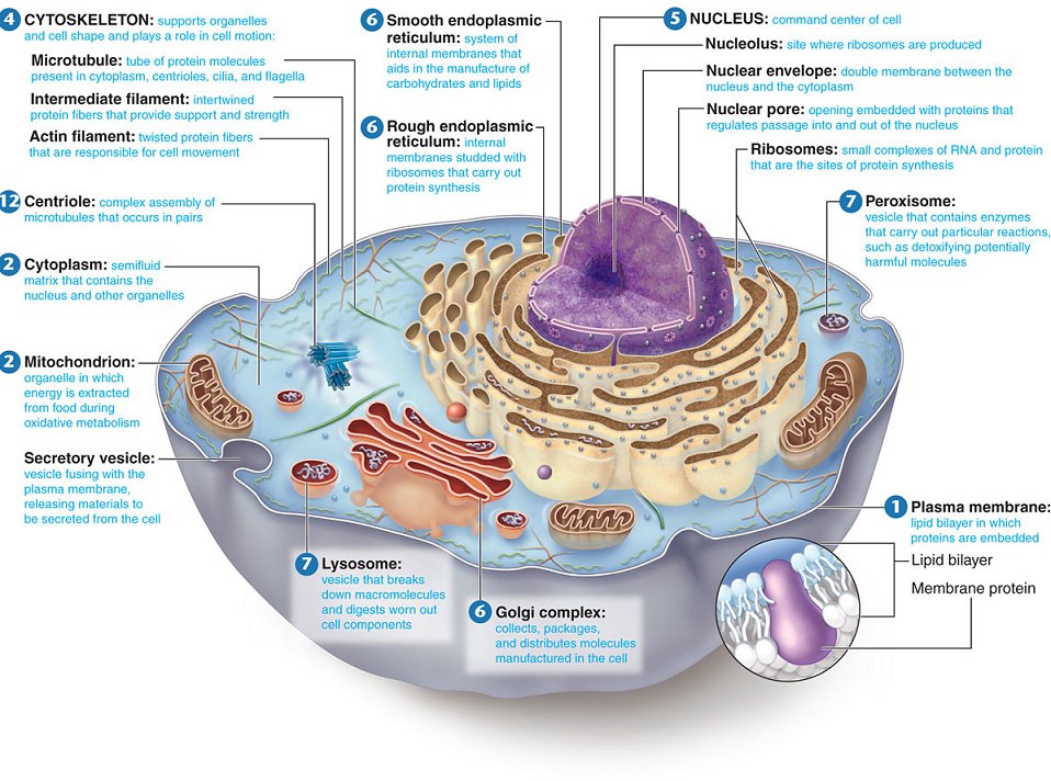
Mitochondria: These double membrane-bound organelles are the tiny powerhouses of the cell, producing ATP (adesnosine-5’- triphosphate), a nucleotide coenzyme that transports energy for use within the cell.
Cytoplasm: The inside of the cell, between the nucleus and plasma membrane, is filled with a gel-like fluid in which the organelles are suspended. The liquid portion of cytoplasm is called cytosol.
Cytoskeleton: This network of fibers and tubules is present throughout the interior of the cell, providing support, anchoring organelles, helping with intracellular transport and cell division.
Centrioles and centrosomes: Only present in animal cells and some fungal cells, a pair of centrioles is located near the nucleus, in a region called the centrosome. These organelles are composed of microtubules, help build flagella and cilia, and appear to be involved in cell division.
Sources & Helpful Cell Biology Links
Bauman, R. (2005) Microbiology. Pearson Benjamin Cummings.
Becker, W. M. et. al. (2009) The World of the Cell. Pearson Benjamin Cummings.
Starr, C. & Taggart, R. (1992) Biology: The Unity and Diversity of Life. Wadsworth Publishing.
Interactive Animal Cell Model from Cells Alive
Inside a Cell, interactive cell diagram from University of Utah
Endoplasmic Reticulum & Golgi Apparatus interactive lesson
Cell Structure Interactive Animation from Wiley.com
Interactive Plant and Animal Cell Simulation by ForgeFX Simulations
Eukaryotic Cell Structure & Function Lecture Main Page, Virtual Cell Biology Classroom
http://en.wikipedia.org/wiki/File:Endomembrane_system_diagram_en.svg
Membranous Organelles of Animal Cells
All membranous organelles are enclosed by the same type of material as the plasma membrane, and can therefore ship materials to each other, via the endomembrane system, in which a piece of a membrane-bound organelle breaks off from one organelle, travels, and then fuses with a different membrane-bound organelle. Material can also enter (endocytosis) or exit (exocytosis) the cell via this method.
Nucleus: Often the largest and most visible organelle in an animal cell, the nucleus is bound by a double-layer nuclear membrane and contains the genetic material (the genome) of the cell. It is also filled with fluid, called nucleoplasm and may contain one or more nucleoli (regions where ribonucleic acid or RNA is synthesized).
Endoplasmic reticulum: A network of hollow tubes, called the endoplasmic reticulum (ER), extends off the nuclear membrane. There are two types of ER. One is rough endoplasmic reticulum, which is studded with ribosomes and involved in making and shipping proteins. Ribosomes are the protein making machinery of the cell, and cells contain many thousands of these tiny nonmembrane-bound organelles. Some ribosomes are attached to the rough ER, others float freely in the cell. The other type of ER, called smooth endoplasmic reticulum, does not have ribosomes, and is involved in the synthesis and transport of lipids.
Vesicles: These small shipping organelles are like membrane bubbles that break off from plasma membrane or membrane-bound organelles and ship materials into, out of or within the cell.
Lysosomes: This special type of vesicle contains lysozyme—an enzyme that can break down organic materials. Lysosomes are produced by the Golgi apparatus and function in cellular digestion and recycling of particles and broken organelles.
Peroxisomes: These vesicles, derived from the endoplasmic reticulum, contain enzymes, such as oxidase and catalase, that break down dangerous free radicals and hydrogen peroxide. Peroxisomes are required by any cell that uses aerobic respiration (oxygen to extract energy from food).
Golgi body: In addition to making lysosmes, this organelle coordinates the packaging and shipment of materials out of the cell.
Stained Volvox colonies, a type of green algae magnified @ 100xTMStained Volvox colonies, a type of green algae magnified @ 100xTM.Stained Volvox colonies, a type of green algae magnified @ 100xTM.
Stained Volvox colonies, a type of green algae magnified @ 100xTM.
facebook link
SPO twitter
Tamislinked in page
SPO VIRTUAL CLASSROOMS
PayPal - The safer, easier way to pay online!
SPO is a FREE science education website. Donations are key in helping us provide this resource with fewer ads.
Please help!
(This donation link uses PayPal on a secure connection.)
Portions of this article originally appeared on Suite101 online magazine.
Page last updated 5/2013
SPO VIDEO: How to Make a Wet Mount of Stained Epithelial Cheek Cells
Instructor's Corner
Cell Envelope & External Structures of Animal Cells
Glycocalyx: Some animal and protozoan cells have this sticky outer layer anchored to the plasma membrane. Gycocalyces help animal cells stick to each other and protect cells from dehydration. This layer is not present in cells that have a cell wall, such as plants, algae and fungi.
Cilia and flagella: Constructed of microtubules covered with plasma membrane (analogous to an arm or leg bone covered in skin), these external appendages, present in some animal cells, aid in cell movement and in moving materials around the outside surface of the cell.
Plasma membrane: All cells have plasma membranes. In eukaryotic cells, this barrier between the inside and the outside of the cell is made
Animal Cell Functions Animal Cell Model Diagram Project Parts Structure Labeled Coloring and Plant Cell Organelles Cake

Animal Cell Functions Animal Cell Model Diagram Project Parts Structure Labeled Coloring and Plant Cell Organelles Cake
Animal Cell Functions Animal Cell Model Diagram Project Parts Structure Labeled Coloring and Plant Cell Organelles Cake

Animal Cell Functions Animal Cell Model Diagram Project Parts Structure Labeled Coloring and Plant Cell Organelles Cake

Animal Cell Functions Animal Cell Model Diagram Project Parts Structure Labeled Coloring and Plant Cell Organelles Cake

Animal Cell Functions Animal Cell Model Diagram Project Parts Structure Labeled Coloring and Plant Cell Organelles Cake

Animal Cell Functions Animal Cell Model Diagram Project Parts Structure Labeled Coloring and Plant Cell Organelles Cake

Animal Cell Functions Animal Cell Model Diagram Project Parts Structure Labeled Coloring and Plant Cell Organelles Cake

Animal Cell Functions Animal Cell Model Diagram Project Parts Structure Labeled Coloring and Plant Cell Organelles Cake

Animal Cell Functions Animal Cell Model Diagram Project Parts Structure Labeled Coloring and Plant Cell Organelles Cake

Animal Cell Functions Animal Cell Model Diagram Project Parts Structure Labeled Coloring and Plant Cell Organelles Cake
nice
ReplyDelete