Animal Cell Anatomy Biography
Source:- Google.com.pk
After completing this section, you should know:
that cells can be different shapes and sizes
the role and function of the plasma membrane; cytoplasm, ribosomes, rough endoplasmic reticulum; smooth endoplasmic reticulum, mitochondria, golgi bodies, lysosomes, centrioles and the nucleus
the structure of the plasma membrane
that substances move across the plasma membrane by passive and active processes
that passive processes include diffusion, osmosis and facilitated diffusion and active processes include active transport, pinocytosis, phagocytosis and exocytosis
what the terms hypotonic, hypertonic isotonic and haemolysis mean
that the nucleus contains the chromosomes formed from DNA
that mitosis is the means by which ordinary cells divide
the main stages of mitosis
that meiosis is the process by which the chromosome number is halved when ova and sperm are formed
The Cell[edit]
Diagram 3.1: A variety of animal cells
The cell is the basic building block of living organisms. Bacteria and the parasite that causes malaria consist of single cells, while plants and animals are made up of trillions of cells. Most cells are spherical or cube shaped but some are a range of different shapes (see diagram 3.1).
Most cells are so small that a microscope is needed to see them, although a few cells, e.g. the ostrich’s egg, are so large that they could make a meal for several people.
A normal cell is about 0.02 of a millimetre (0.02mm) in diameter. (Small distances like this are normally expressed in micrometres or microns (μm). Note there are 1000 μms in every mm).
Diagram 3.2: An animal cell
When you look at a typical animal cell with a light microscope it seems quite simple with only a few structures visible (see diagram 3.2).
Three main parts can be seen:
an outer cell wall or plasma membrane,
an inner region called the cytoplasm and
the nucleus
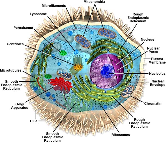
Diagram 3.3: An animal cell as seen with an electron microscope
However, when you use an electron microscope to increase the magnification many thousands of times you see that these seemingly simple structures are incredibly complex, each with its own specialized function. For example the plasma membrane is seen to be a double layer and the cytoplasm contains many special structures called organelles (meaning little organs) which are described below. A drawing of the cell as seen with an electron microscope is shown in diagram 3.3.
The Plasma Membrane[edit]
Diagram 3.4: The structure of the plasma membrane
The thin plasma membrane surrounds the cell, separating its contents from the surroundings and controlling what enters and leaves the cell. The plasma membrane is composed of two main molecules,phospholipids(fats) and proteins. The phospholipids are arranged in a double layer with the large protein molecules dotted about in the membrane (see diagram 3.4). Some of the protein molecules form tiny channels in the membrane while others help transport substances from one side of the membrane to the other.
How substances move across the Plasma Membrane[edit]
Substances need to pass through the membrane to enter or leave the cell and they do so in a number of ways. Some of these processes require no energy i.e. they are passive, while others require energy i.e. they are active.
Passive processes include: a) diffusion and b) osmosis, while active processes include: c) active transport, d) phagocytosis, e) pinocytosis and f) exocytosis. These will be described below.
Diagram 3.5: Diffusion in a liquid
a) Diffusion
Although you may not know it, you are already familiar with the process of diffusion. It is diffusion that causes a smell (expensive perfume or smelly socks) in one part of the room to gradually move through the room so it can be smelt on the other side. Diffusion occurs in the air and in liquids.
Diagram 3.5 shows what happens when a few crystals of a dark purple dye called potassium permanganate are dropped into a beaker of water. The dye molecules diffuse into the water moving from high to low concentrations so they become evenly distributed throughout the beaker.
In the body, diffusion causes molecules that are in a high concentration on one side of the cell membrane to move across the membrane until they are present in equal concentrations on both sides. It takes place because all molecules have an in-built vibration that causes them to move and collide until they are evenly distributed. It is an absolutely natural process that requires no added energy.
Small molecules like oxygen, carbon dioxide, water and ammonia as well as fats, diffuse directly through the double fat layer of the membrane. The small molecules named above as well as a variety of charged particles (ions) also diffuse through the protein-lined channels. Larger molecules like glucose attach to a carrier molecule that aids their diffusion through the membrane. This is called facilitated diffusion.
In the animal’s body diffusion is important for moving oxygen and carbon dioxide between the lungs and the blood, for moving digested food molecules from the gut into the blood and for the removal of waste products from the cell.
Diagram 3.6: Osmosis
b) Osmosis
Although the word may be unfamiliar, you are almost certainly acquainted with the effects of osmosis. It is osmosis that plumps out dried fruit when you soak it before making a fruit cake or makes that wizened old carrot look almost like new when you soak it in water. Osmosis is in fact the diffusion of water across a membrane that allows water across but not larger molecules. This kind of membrane is called a semi-permeable membrane.
Take a look at side A of diagram 3.6. It shows a container divided into two parts by an artificial semi-permeable membrane. Water is poured into one part while a solution containing salt is poured into the other part. Water can cross the membrane but the salt cannot. The water crosses the semi-permeable membrane by diffusion until there is an equal amount of water on both sides of the membrane. The effect of this would be to make the salt solution more diluted and cause the level of the liquid in the right-hand side of the container to rise so it looked like side B of diagram 3.6. This movement of water across the semi-permeable membrane is called osmosis. It is a completely natural process that requires no outside energy.
Although it would be difficult to do in practice, imagine that you could now take a plunger and push down on the fluid in the right-hand side of container B so that it flowed back across the semi-permeable membrane until the level of fluid on both sides was equal again. If you could measure the pressure required to do this, this would be equal to the osmotic pressure of the salt solution. (This is a rather advanced concept at this stage but you will meet this term again when you study fluid balance later in the course).
Diagram 3.7: Osmosis in red cells placed in a hypotonic solution
The plasma membrane of cells acts as a semi-permeable membrane. If red blood cells, for example, are placed in water, the water crosses the membrane to make the amount of water on both sides of it equal (see diagram 3.7). This means that the water moves into the cell causing it to swell. This can occur to such an extent that the cell actually bursts to release its contents. This bursting of red blood cells is called haemolysis. In a situation such as this when the solution on one side of a semi-permeable membrane has a lower concentration than that on the other side, the first solution is said to be hypotonic to the second.
Diagram 3.8: Osmosis in red cells placed in a hypertonic solution
Now think what would happen if red blood cells were placed in a salt solution that has a higher salt concentration than the solution within the cells (see diagram 3.8). Such a bathing solution is called a hypertonic solution. In this situation the “concentration” of water within the cells would be higher than that outside the cells. Osmosis (diffusion of water) would then occur from the inside of the cells to the outside solution, causing the cells to shrink.
Diagram 3.9: Red cells placed in an isotonic solution
A solution that contains 0.9% salt has the same concentration as body fluids and the solution within red cells. Cells placed in such a solution would neither swell nor shrink (see diagram 3.9). This solution is called an isotonic solution. This strength of salt solution is often called normal saline and is used when replacing an animal’s body fluids or when cells like red blood cells have to be suspended in fluid.
Remember - osmosis is a special kind of diffusion. It is the diffusion of water molecules across a semi-permeable membrane. It is a completely passive process and requires no energy.
Sometimes it is difficult to remember which way the water molecules move. Although it is not strictly true in a biological sense, many students use the phrase “SALT SUCKS” to help them remember which way water moves across the membrane when there are two solutions of different salt concentrations on either side.
Animal Cell Anatomy Animal Cell Model Diagram Project Parts Structure Labeled Coloring and Plant Cell Organelles Cake
Animal Cell Anatomy Animal Cell Model Diagram Project Parts Structure Labeled Coloring and Plant Cell Organelles Cake

Animal Cell Anatomy Animal Cell Model Diagram Project Parts Structure Labeled Coloring and Plant Cell Organelles Cake

Animal Cell Anatomy Animal Cell Model Diagram Project Parts Structure Labeled Coloring and Plant Cell Organelles Cake

Animal Cell Anatomy Animal Cell Model Diagram Project Parts Structure Labeled Coloring and Plant Cell Organelles Cake
Animal Cell Anatomy Animal Cell Model Diagram Project Parts Structure Labeled Coloring and Plant Cell Organelles Cake

Animal Cell Anatomy Animal Cell Model Diagram Project Parts Structure Labeled Coloring and Plant Cell Organelles Cake

Animal Cell Anatomy Animal Cell Model Diagram Project Parts Structure Labeled Coloring and Plant Cell Organelles Cake
Animal Cell Anatomy Animal Cell Model Diagram Project Parts Structure Labeled Coloring and Plant Cell Organelles Cake

Animal Cell Anatomy Animal Cell Model Diagram Project Parts Structure Labeled Coloring and Plant Cell Organelles Cake

Animal Cell Anatomy Animal Cell Model Diagram Project Parts Structure Labeled Coloring and Plant Cell Organelles Cake
No comments:
Post a Comment